Friday, August 3, 2007

Neurological Surgery

Neurological Surgery

During the course of this program, I have had the opportunity observe several different treatments for the same illness. I was able to observe a carotid endarterectomy and brain tumor resection, which were particularly interesting because I have observed the alternative endovascular treatments for each, carotid artery stenting (CAS) and embolizing the vessels feeding the tumors. Though I have written about the advantages of CAS over endarterectomies in the past, I was able to directly observe the vast improvement that CAS provides in terms of time and invasiveness - unlike in carotid endarterectomies where blood flow is cut off through one of the carotid arteries that feeds the brain for close to 40 minutes, CAS does not require blood flow to be cut off at all. Similarly, the endovascular method to deprive tumors of blood is much less invasive than the surgical technique which requires a craniotomy. In the case I observed, the doctors had to be very careful about avoiding damage to a major artery and the optic nerve while removing the tumor. In terms of biomedical engineering contributions, technology that could decrease invasiveness and improve precision in treatments would be very useful.

I also have been working on my clinical research project evaluating effectiveness of two different treatments for aneurysms, and have obtained some interesting results. I will be presenting my research project in the seminar meeting in Ithaca in the fall.

Overall, I gained a lot from this experience, especially in terms of realizing that there are many areas that still need technological improvements. I would like to thank my clinician mentor, Dr. Riina, and also Dr. Gobin and Dr. Chapple.

Thursday, August 2, 2007

Vascular Wrap-up

Vascular Surgery
Week 7


The Last Post

Last time I left a taunting message about my research—now it’s time for some results! It turned out that the average age of the last menses was 48 and that surgery would occur within 20-29 years after this age with 99% statistical confidence. This identified the patient population with ages 68-77. The HRT data revealed that there was no significant difference between patients on HRT vs. not for primary patency while patients with osteoporosis were worse-off than those without osteoporosis. This is interesting data that may suggest that patients with osteoporosis who undergo vascular procedures should have more frequent patency check-ups.

This is the last post for me! One thing that I pulled away from this experience is that I do not want to be a doctor, at least not a surgeon! They have an intense lifestyle to say the least, and they basically live at the hospital. Otherwise I had a chance to see some great technology and procedures that I wouldn’t be exposed to anywhere else, and living on the Upper East Side rent-free was unbeatable. Thanks to Dr. Vouyouka, and thanks for reading!

Surgery and Adios

This past week I spent time observing thoracic surgery. This was the first time I saw surgery, as I found other topics more interesting/relevant for me, my research, and my personal goals for the program. However, one surgery in particular was very interesting. It was a bi-lobectomy. In this surgery, the 2 lower lobes from the right lung were removed. As a note, the right lung has 3 lobes, the left two (because the heart is in the way. The patient had lung cancer and resection (removal) was deemed to be the method with the highest chance of success. The reason two lobes were removed is because it was felt the cancer was large enough and was invading/compromising the second lobe. What I found interesting was that this decision was made in the OR and not before hand, which shows the adaptive nature of the OR. I did have to get used to the cauterization though. It looked like they were soldering the patient, and it threw me the first time I saw it.

The summer immersion program is almost over now, and I think that I did gain a better understanding of clinical practice. I am fairly sure this experience will spill over into my current collaborations, and will benefit me. It also gave me a better understanding of the need to be assertive. The programs structure does need refinement, however, but it is not a loss, especially if one tries to gain specific insight on one’s own. It would have been nice if the exact clinical experiences we were expected to have (and it did feel as if there explicit expectations) would have been spelled out clearly before hand.

I’m looking forward to returning home to my wife and child tomorrow. Happy 1st birthday, Aiden!

Mitral Valve Replacement (MVR)

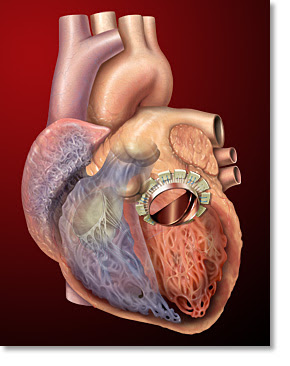
Finally, I convinced myself to watch at least one open heart surgery. I was surprised at how invasive the whole procedure was. The doctors’ basically sedated the patient, sliced his chest open, and then pulled his ribs apart using their bare hands or a crude mechanical device. It was almost like watching a scene out of a movie with hygienic zombies in scrubs trying to carefully eat the victim’s heart. Okay, maybe that’s an exaggeration. What followed was a bit more impressive. They bypassed the patient’s arteries, immobilized the heart using high concentration of potassium chloride, and then made an incision in the heart in order to replace the mitral valve. The following picture is borrowed from its.med.yale.edu. It depicts a mitral valve replacement.Apparently, this particular patient had mitral valve regurgitation. This was evident from his transesophogeal echocardiogram or the ultrasound measurements. One could see that the oxygenated blood that entered the left ventricle through the mitral valve periodically reentered the left atrium. In most cases, a patient’s valve is irreversibly compromised due to either ischemia, stenosis, or infection. These problems are usually onset due to coronary artery disease, age, or rheumatic fever, respectively. However, congenital defects are not rare.

In the patient’s case, the problem was caused by ischemia through coronary artery disease. As a result, the doctor had decided to replace his valve with a bovine valve - the reasons for choosing a bovine valve or a mechanical valve were explained previously by Dickinson. It seemed as though the stitching of the valve required great dexterity. One of the more skilled residents tried to stitch the valve but it seemed as though he could not make the more difficult 10-12 o’clock stitches. As a result, the primary surgeon took over and showed him how it should be done.

One could definitely make it easier for these doctors by developing an innovative device for sewing. In fact, I don’t really know why a big hospital like NYP still uses open heart surgery for such cases. There are hospitals that are performing these types of procedures percutaneously using the da Vinci surgical system. I think that’s the way to go. If not, the least one should do is make something that replaces the “needle and string.”

My Last Week in Plastics

This last week has been very eventful. On Monday at the residents discussion the talk was on Breast Reductions.
First the discussion was on different types of reductions, mainly different types of incisions and techniques used to reduce the breast size. Then there was a good discussion of how to decide what type of reduction the patient will receive. Specific cases are discussed and the idea of what is realistic and what is not was gone over. Many of the scars after the surgeries are quite noticeable. Images from : www.aboardcertifiedplasticsurgeonresource.com.
Generally, an incision is made around the nipple and the nipple is then removed and a new nipple relocation is added to raise the nipple site to fit with the new smaller breast. Tissue is removed along with skin and then brought in downward to close over and shape the new breast. Scars will remain around the nipple, extended to the crease, and through the crease as seen in the pictures. Later that day, a patient came to office hours to be consulted for a breast reduction. Mistakes were also discussed where the reduction produced breasts that were very asymmetric and occasionally with nipples that were misplaced. Many of these complications arise because the doctor works with an assistant on one side or because of shifts from laying on teh operating bed to getting up. This really brought the talk together because I went from the academic discussion to the patient doctor interactions.

Additionally, I attended the M&M meeting Monday evening were complications for the past month in the plastics department were discussed. No patients died of complications, but additionally surgeries were required. In one case a tissue expander became infected after only a week and after taking intravenous antibiotics the patient opted to have the expander removed.

At office hours I was able to the progress of the patient I spoke about last entry and the V.A.C. has continued to help in wound closure progress. I also helped to remove sutures. The patient had a cut that ran down the side of the face and a second cut on the upper back. A running stitch was used to close the face face wound.

I also attended a butt flap surgery where a patient had gotten a bed ulcer after lying on their back for an extended amount of time. In order to close the wound the muscle above the wound was mobilized and swung down to close over the wound.

I have also spent time working on the Case Report that I am writing up. I have submitted a draft to the Chief Resident that I am working with to get feedback on format and wording.

Code Blue in the OR

During this week I was able to observe a few more surgeries and attend a few more rounds. I was luckily able to scrub into another tracheaesophageal fistula. The patient was two days old and had a type C fistula which consists of an upper esophagus ending in a blind pouch and a connection between trachea and fistula. Dr. Spigland was able to occlude the fistula and connect the esophagus back together.

However, during the esophageoesophagus connection the patient’s lung did not infant. The lung appear very deflated and small. A code blue was called and within 1-2 minutes a barrage of nurses, residents, and attendings came to help out. They ran into the room with concern expressions and eagerness to help out. The attendees quickly got the patient to start breathing again using manual ventilation. There was a pediatric cart in the room in case the use of a defiberator was needed. In actuality, most of the people who rushed in for the code blue just stood around watching while 1-2 attendees did all the work. But it’s a good sign when over a dozen people rush into the room minutes after a code was called.

I was able to make progress on my research and conclude some aspects of the project. I will continue to collaborate with Dr. Spigland on the research project dealing with esophageal atresia and hopefully write something up in the near future.

Wednesday, August 1, 2007

Calcium Score: Part 2

Okay… I think I promised you that I would upload something on a new GE software, which I was supposed to use this past week to measure calcium score. Apparently, there are some legal issues in distributing the software so I don’t think I’ll be using it before we all leave for Ithaca. In a perfect world, I would have compared the calcium score measurements made by Smart Score (discussed last time) to those made by VCAR (the new GE software). Instead, I will only have the Smart Score measurements done and my mentor, Dr. Min, will finish the VCAR measurements whenever he gets the software. I think I will still get my name on a paper since he plans to send out a manuscript highlighting the results of the comparison.

Maybe it is better in a certain way that I will not be collecting scores using VCAR. According to my mentor, although the software is more advanced than Smart Score, it is just as tedious to use. If you recall, the Smart Score software forced one to highlight the calcium in transverse CTA slices. It, thereby, was able to deduce a calcium score based on just volume (volumetric score) or area and average Hounsfield value (agatson number) for the overall calcium in the arteries. VCAR calculates the score in a similar fashion, but it does not need the user to highlight the calcium. Instead, it is designed to automatically segment the calcium in the arteries. However, before the algorithm is capable of doing this, one must click along the centerline of an artery so that the algorithm can basically fit a curve to the selected points and subsequently subtract it from the image to straighten out the artery. Then it presents the artery of interest in a longitudinal format so that one is better able to view the calcium deposits (figure). I’m not certain why this step is necessary. Nevertheless, the software seems very user friendly so I think it would have been fun to work with.