Monday, July 16, 2007

As with anything, there are no guarantees...

This past week (7/9-7/13) I had the privilige of going on rounds with my clinician and meeting patients. It was the first time I got to see what problems the patient had and how the doctor planned to fix it. Previously, I had been mainly observing surgeries in the OR, in which we really don't see the evolution of the treatment, just the actual "fixing." One of the first patients I got to meet suffered from partial seizures. There are two main types of seizures: partial and generalized. In partial seizures, the most common type, the electrical activity is limited to one focal area in the brain. In generalized seizures, the electrical activity propagates throughout both hemispheres of the brain and can be usually characterized by loss of conciousness. This patient suffered from numbness of the left side of her body and prior to the onset of a seizure, would feel great anxiety and experience an aura of some sort. She had been on anti-epileptic drugs, but they were no longer effective. The cause of her seizures were rooted in the tumor she had. She had previously had surgery to remove the tumoregenic tissue and to determine the state of the tumor. Now the tumor had grown back and making her seizures worse. To treat her, the best option would be to remove the tissue. However, the tumor was in a sensitive area where some crucial motor neurons were still intact. She was told that surgery would most likely improve the frequency and level of her epileptic episodes, but there were no guarantees and that she would still most likely experience the numbness in the left side of her body.


Another patient we saw, had a tumor that grew fives time its size from 5 years ago. The figure above shows an MRI scan of a brain tumor in the brain. Here the tumor looks white. This is becuase the tumoregenic tissue produces high contrast relative to normal braint tissue. This patient had done extensive research and knew what he was getting into. What was different about this case was that his family was in the room with him. It was a very intense situation for me, to see how the family was reacting to the prognosis of his condition. No matter what numbers my clinician was giving him, he didn't seem to understand that nothing is guaranteed. I believe he was going to get back to my clinician on what he planned on doing. One of the more interesting aspects of this case was how adamant the patient was on getting the statistics of his outcome. To me, the prognosis was obvious: you have a huge tumor in your head, take it out or else you will definitely lose cognitive function. Pecentages are all relative.

Neuro Detour

Vascular Surgery
Week 4


This week I took a detour from vascular surgery to watch a couple of neurological surgery cases. One really interesting case was the removal of a pituitary tumor.

Removal of Pituitary Tumor
Endoscopic Endonasal Surgery


http://www.skullbaseinstitute.com/video_pituitary_tumor.htm

There is a great video of the procedure at the link above. A few stills are shown below:


The endoscope is inserted through the nostril and navigated to the brain.

The dura (covering of the brain) is incised.

The tumor is gradually removed.

Source: http://www.skullbaseinstitute.com/video_pituitary_tumor.htm

Research Project

I’ve also been working on my research project regarding the outcomes of vascular procedures in women. Some of the demographics of the telephone interviews I have been conducting are shown below:

This chart shows the proportions of women I have contacted that are on or have had hormone replacement therapy (HRT), have been diagnosed with osteoporosis, take osteoporosis supplements, and who take calcium or vitamin supplements (total does not equal 100%). I’m still working on the data analysis regarding the outcomes of procedures and hope to have results soon!

Sunday, July 15, 2007

From Research to the Real World

As I mentioned in my previous post, my research topic is on esophageal atresia (EA) with or without tracheoesophageal fistula (TEF). The disease is a congenital underdevelopment of the esophagus and trachea. These two tubes, for the most part, can be connected to each other, making eating and breathing almost impossible for the patient. The disease is not rare among newborns and has a prevalence of about 1 in 4000. For the past few weeks, I have just been reading up and doing statistical analysis on past cases on EA/TEF. To my surprise there happened to be a patient born and admitted in the neonatal intensive care unit (NICU) a few weeks ago with EA/TEF. I took the time to check out the patient and the patient history as my research and the real world were finally integrated.

umd.edu

The patient was born with the most common type EA/TEF. Her upper esophagus discontinued into a pouch and her bottom esophagus connected to her trachea (which would be the fistula part). As of recent decades, this repair would be an easy fix with a low mortality rate. Normally, the patient would be operated on within the first 48 hours, which is known as primary repair. However, this patient, and like 30% of all EA/TEF patients, has some type of other complication. In this case it was cardiac problems. The patient had to be admitted into surgery to fix her heart problem before fixing the EA/TEF. She would not be able to survive the EA/TEF recovery without everything else being healthy.

I wasn’t there for her heart operation, but found out that when the doctors closed the patients chest back up, the heart was beating against the chest wall causing too much pressure. The doctors, then had to re-open the chest and leave it open for a few days until she stabilized from heart sugery. They used some type of plastic see-thru-wrap (much like cling-wrap) to close up the chest cavity from the outside environment. This allowed the heart to beat freely. She remained in the NICU until her body stabilized; upon stabilization she returned to the operating room to have her chest closed. She will be back to the operating room in a few days (weeks?) to have her esophagus disconnected from her trachea and to connect the two disconnected esophagus’. They are waiting for her to stabilize before performing this operation.

In retrospect, the complications the patient had with her EA/TEF would have led to a low survival rate only 10-20 some years ago. Over the past decade the overall mortality for EA/TEF have gone down to 11% from 22%. This is due to refinements in NICU care, anesthetic management, ventilator support, and improved surgical techniques. The corrective surgery for EA/TEF is at a 96-98% success rate presently, but was at a 0% survival rate just 50 years ago.

As researchers especially in academic research, we do a lot of basic science –discovering this, studying about that. A lot of our research won’t make a difference in the real world until decades from now. This other realm of research I’ve been exposed to has some gratifying feelings, that being once you’re done with your research, it will have a direct impact on people and their lives.

4th Week in Plastics

I started off the past week with a Monday morning lecture on lower extremity wounds. Dr. Spector gave the lecture which was followed by case reviews for the residents. This was very interesting to watch because the reviews allowed insight into the entire diagnosis and treatment process; describing from when a patient presents with a particular case, noting what knowledge is important to gain about the patient in order to make a proper decision, and finally how to treat the patient and what should be done when obstacles are confronted.

During office hours I saw a burn patient that Dr. Spector has been treating. The patient has scaring all over his face and upper torso which is disfiguring, painful, and prevents proper function. Around his mouth and neck the scarring has hardened so that he is not able to fully open is mouth or rotate his head. In addition, his nose is no longer symmetric which Dr. Spector plans to alter by removing cartilage and skin from his nose to reform the nostril. In addition, I saw quite a few cosmetic consults during office hours where Dr. Spector discusses the desired alterations a patient may want to his or her body including breast augmentations, rhinoplastys, or liposuction. I also met a patient that Dr. Spector performed a breast augmentation for a few years ago and recently one breast has enlarged and become painful as the fibrous tissue scarring has begun to contract. This is a known complication to breast augmentation. In order to fix the problem, the patient will have to undergo surgery to remove the fibrous tissue.

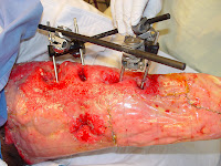
I have also seen some interesting cases in the OR. Prior to my arrival, a patient was admitted after being struck by a vehicle. The resultant injuries required that her femur be externally fixed as seen in the images below to allow for proper repair. Once the internal injuries were stabilized, a V.A.C. system was used to allow the wound to begin to close and create the proper surface on which a skin graft could be placed. Once the leg had healed additionally, Integra ® Dermal Regeneration Template was placed over the wound as seen
in the next image. This is a product made from glucosaminoglycan and collagen which allows for the in growth of blood vessels and provides a better layer on which a skin graft can be added. Once the under lying dermal layers were present, a skin graft was taken from the right leg to cover the wounds on the left leg as seen in the images. The donor sites are left without the outermost skin layers which are usually able to regenerate and heal although a pigment difference may make the donor location visible.


This Week in Plastics - Keloids, VAC

For the past holiday week the OR schedule has been lighter than usual as many of the surgeons have taken a vacation. Most of the procedures I have seen have been debridements or skin grafts. During the skin graft procedure, a very thin layer of skin, which may contain either part or all of the dermis (~15/1000 in thick) is removed from a donor site and cut to fit over the wound. In order for the skin graft to take, the underlying tissue must be healthy, vascularized, and granulated which is often achieved with the V.A.C. machine that I spoke about last week.


In addition to the OR, I have been able to see many very interesting cases during office hours. The nurse that works with Dr. Spector in office hours has shown me where many of the instruments and supplies are stored allowing me to assist Dr. Spector and be more involved with the care of the patient. One patient that has been coming in twice a week for V.A.C. changes has a very persistent wound. The patient had a hernia repair which left a large opening in his abdominal area. A hernia occurs when underlying tissue protrudes through tissue that usually encloses it (generally muscle). As in the diagram the intestines may break through the abdominal muscle wall and require repair (picture from: http://www.pbennett.com.au/images/abdominal_hernia.jpg). In the picture on the right, you can see the black sponge that has been placed over the open wound of a patient that is used with the V.A.C. system to aid in closure (picture from: http://www.emedicine.com/med/images/4884med3722-30 .jpg). The V.A.C. has helped to close the wound, but at a very slow rate and each week during office hours Dr. Spector debrides the wound of dead tissues. It is interesting to see the progress of the patient’s wound closure with the help of a simple suction machine. Many of the cases during office hours are post-operative check-ups, but occasionally there are pre-operative consults.

In addition to consults, Dr. Spector also performs minor procedures during office hours. A patient last week that had a severe case a keloids was treated with steroid injections. Keloids are collections of scar tissue that form after an injury or surgery. Keloids are cosmetically unappealing and are often quiet painful. In the picture below, you can see an example of a keloid (picture from: http://fig.cox.miami.edu/~cmallery/150/protein/keloid.jpg). In this case the keloids had formed after a spinal surgery which meant they were in a very uncomfortable position. Two treatments for keloids that show promise are compression and steroid injections. It is not completely clear how the steroids aid in smoothing out the keloids, but with the injections the patient can see definite progress in both the appearance and pain level of the keloids.

Learning to Tie Sutures

Plastic surgery is essentially there to make you look pretty (…or at least less ugly). As such, wound closure and healing is of utmost importance in order to minimize the effects of scarring. As with most things in life, there are many factors influencing this…one being the surgeon’s hand.

The other day it was relatively slow all around and so I was fortunate enough to sit down with some doctors and doctors-to-be and learned the subtle art of suturing. I’m sure we have all witnessed trained surgeons throwing knots like there’s no tomorrow; however, rather surprisingly there is a lot that goes into even a simply stitch.

So apparently, there are over 1400 different types of knots (or at least according to the Encyclopedia of Knots…who knew there was even such an encyclopedia…shows I was never Eagle Scout extraordinaire). Anyway, despite the plethora of knots, surgeons only use several different ones. The most common are as follows: square knot, surgeon’s knot, deep tie,
instrument tie, running stitch and ligation with a hemostat (a surgical clamp).

The square knot is essentially the same thing as the knot you use to tie your shoelaces, with the exception that, like everything in medicine, you have to make it more complicated than necessary. Similarly, the surgeon’s knot is essentially a double knot. The deep tie is usually used to approximate tissues deep in a cavity. The instrument tie is a quick way to tie a knot by simply wrapping the suture around the needledriver and then using needledriver to pull the other end of the suture through the loop you just made. Both square and surgeon’s knots can be thrown in this manner. The running stitch is just a continuous series of stitches thrown with one suture. I didn’t learn how to do a ligation tie, but I’ve seen it done a few times. It seems like you clamp down a vessel, loop the suture around the hemostat and tie off the vessel behind the clamp with some square knots.

Anyway, this post was not meant to reveal anything profound, I just thought it was an interesting experience to share and some potentially useful knowledge to know…next time I need to tie off my blood vessels I will know…and knowing is half the battle…

Phase Contrast MRI 2: The Revenge

Last week, I talked briefly about how we use successive slices in a cine MRI sequence to evaluate heart function. Using this type of image analysis, we are able to find information such as cardiac output and ejection fraction, as well as volume and mass information during various stages of the heart. In addition, we can use a technique called Phase Contrast MRI to obtain flow information. While the data obtained from Phase Contrast MRI is slightly different than the data obtained from segmenting a cine MRI, it is possible to correlate the two and from this comparison, we can determine the agreement between our two measurements.

Phase contrast works by exploiting the nature of MR imaging. Two gradients, with equal magnitude but opposite orientation, are applied to the area of interest in rapid succession. Objects that remain completely stationary undergo no net phase shift, as the opposing gradients “cancel out” the effects of one another. However, objects that are moving undergo a change in phase proportional to their velocity. This concept is illustrated in the diagram below.


Phase Contrast gradient fields on stationary and moving objects
(image obtained from radiographics.rsnajnls.org)


The top panels demonstrate the gradients on a stationary object. The first gradient “tips” the atoms by varying amounts, depending on their location within the gradient field. These tip angles are eliminated upon the application of the second gradient field. The end result gives the effect as if no gradient was applied at all, and all magnetic moments are uniform and in a single direction.


On the other hand, a moving object will undergo a predictable change in phase. The bottom panels illustrate what happens when a single atom travels linearly through the same region. The end result is a phase distinctly different than that of the stationary atoms.

This information can be visualized by viewing the phase information of a particular MR image sequence. Typically, a neutral gray color is assigned to pixels in which there is no net phase difference. Black indicates that the objects within the pixel are moving towards the viewer, while a white color indicates that objects are moving away. Varying velocities are indicated by the intensity of these shapes. From this, we can quickly determine the direction and amount of flow. Further, by using computer automation and manual segmentation, we can determine the amount of flow in specific areas.

In this study, we will be performing phase contrast analyses of the aorta. By quantifying the amount of blood flowing through the aorta, we can indirectly determine the stroke volume of the heart, which will provide an additional form of “ground truth” by which we can further evaluate our automatic segmentation algorithm.


Magnitude MR image ("Regular")
(image obtained from radiographics.rsnajnls.org)


Phase Contrast MR image (new and improved!)
(image obtained from radiographics.rsnajnls.org)